Home » Uncategories » Nasal Bone Anatomy Ct Radiology : Technology and Techniques in Radiology: CT Anatomy of the ... : If you sustain a nasal injury, it is best to be examined by a doctor.
Nasal Bone Anatomy Ct Radiology : Technology and Techniques in Radiology: CT Anatomy of the ... : If you sustain a nasal injury, it is best to be examined by a doctor.
Nasal Bone Anatomy Ct Radiology : Technology and Techniques in Radiology: CT Anatomy of the ... : If you sustain a nasal injury, it is best to be examined by a doctor.. Intended for beginning radiology residents, this video highlights important structures in the temporal bone to evaluate on every temporal bone ct. Learn about the anatomy of the skull bones and sutures as seen on ct images of the brain. Gross anatomy the nasal bone has two surfaces: Understanding the anatomy of the temporal bone has always been, is and will be a difcult task for doctors of various specialities: Limson (1932) commented on the variety in size and shape of fetal nasal bones and distinguished four distinct classes according to overall shape, the percentage frequencies being.
If you sustain a nasal injury, it is best to be examined by a doctor. If we are not able to know the level yet, we can use some bone structures to identifyit. To know what is what, first you should remember their anatomical location in order to know where to expect them; Here we will discuss the anatomy of the nasal skeleton and its component bones. Pictures show possible high fracture of right side better appreciated as compared to the left, though airzones and soft tissues are not grossly abnormal.
High-resolution thin-section CT scan of the skull base ... from www.researchgate.net To know what is what, first you should remember their anatomical location in order to know where to expect them; Intended for beginning radiology residents, this video highlights important structures in the temporal bone to evaluate on every temporal bone ct. The nasal bones are two oblong halves that form the roof of the bony vault of your nose. Learn about the anatomy of the skull bones and sutures as seen on ct images of the brain. Posted by radiologypics ⋅ march 21, 2013 ⋅ 1 comment. The anatomy of the ethmoid ct scan, nasal cavity. Basic ct anatomy of paranasal sinuses, made easy in this video you will learn the basic normal anatomy of the nasal cavity and. Overview on what a radiologist is and what they do.
By dr.k.prasanna radiology resident rmmch 2.
Normal anterior ostiomeatal complex anatomy. To help determine the level of the scan we can should try to identify the larger organs such as the liver, the spleen, the kidneys. To load the skull base ct anatomy module in a new window click on its image above. External surface attaches to t. 13.10.2014 · ct anatomy of para nasal sinuses 1. External surface attaches to t. Given that the file is large, loading may take a few minutes. Ct anatomy of the paranasal sinuses by hazem youssef 11485 views. The function of each nasal bone is to bind together the cartilage that forms individual nose contours and shapes. Understanding the anatomy of the temporal bone has always been, is and will be a difcult task for doctors of various specialities: Gross anatomy the nasal bone has two surfaces: The nasal bones develop in membrane in the dense mesenchyme overlying the cartilaginous nasal capsule. Nose and nasal fossa para nasal sinuses osteomeatal complex anatomical variations imaging modalities ct procedure.
If you sustain a nasal injury, it is best to be examined by a doctor. Learn about the anatomy of the skull bones and sutures as seen on ct images of the brain. In their first year, residents should be well versed with normal radiographs, ultrasound and ct anatomy followed by mri in the consequent years. its ability to optimally display bone. Gross anatomy the nasal bone has two surfaces:
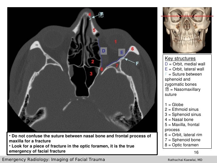
Imaging Of Facial Trauma Part 1 from image.slidesharecdn.com Pictures show possible high fracture of right side better appreciated as compared to the left, though airzones and soft tissues are not grossly abnormal. Understanding the anatomy of the temporal bone has always been, is and will be a difcult task for doctors of various specialities: The anatomy of the ethmoid ct scan, nasal cavity. The nasal anatomy shows much individual variation. If we are not able to know the level yet, we can use some bone structures to identifyit. Ct delineates bony abnormalities better than mri, allowing determination of the cribriform plate's level (the bone that transmits the olfactory nerves that. The nasal septum consists of both bone tissue and cartilage. Information about the sinus anatomy of individual patients is essential prior the nose is divided in two by the nasal septum and reaches up to the nasopharynx.
Nasal bones are normally small and oblong, but can differ in size and shape in different people.
Usually from direct blow during athletics, motor vehicle collision or an altercation; Limson (1932) commented on the variety in size and shape of fetal nasal bones and distinguished four distinct classes according to overall shape, the percentage frequencies being. I am a radiology physician from california, usa. External surface attaches to t. The module interface is meant to mimic a radiology workstation with adjacent image scrolling via arrow keys and or mouse wheel button. its ability to optimally display bone. Cross sectional anatomy of paranasa 63. 4, lamina papyracea of the ethmoid bone. Understanding the anatomy of the temporal bone has always been, is and will be a difcult task for doctors of various specialities: Ct delineates bony abnormalities better than mri, allowing determination of the cribriform plate's level (the bone that transmits the olfactory nerves that. The nasal anatomy shows much individual variation. Information about the sinus anatomy of individual patients is essential prior the nose is divided in two by the nasal septum and reaches up to the nasopharynx. To help determine the level of the scan we can should try to identify the larger organs such as the liver, the spleen, the kidneys.
Limson (1932) commented on the variety in size and shape of fetal nasal bones and distinguished four distinct classes according to overall shape, the percentage frequencies being. To know what is what, first you should remember their anatomical location in order to know where to expect them; ct is currently the modality of choice in the evaluation of the paranasal sinuses and adjacent structures. Given that the file is large, loading may take a few minutes. Usually from direct blow during athletics, motor vehicle collision or an altercation;
#Face #CT shows #nasal #fractures (arrow) and nasal # ... from i.pinimg.com To know what is what, first you should remember their anatomical location in order to know where to expect them; its ability to optimally display bone. If we are not able to know the level yet, we can use some bone structures to identifyit. I am a radiology physician from california, usa. The nasal septum consists of both bone tissue and cartilage. External surface attaches to t. Nose and nasal fossa para nasal sinuses osteomeatal complex anatomical variations imaging modalities ct procedure. Here we will discuss the anatomy of the nasal skeleton and its component bones.
The nasal bones are two oblong halves that form the roof of the bony vault of your nose.
To load the skull base ct anatomy module in a new window click on its image above. ct is currently the modality of choice in the evaluation of the paranasal sinuses and adjacent structures. This mri knee cross sectional anatomy tool is absolutely free to use. Overview on what a radiologist is and what they do. Ct delineates bony abnormalities better than mri, allowing determination of the cribriform plate's level (the bone that transmits the olfactory nerves that. Setting up a radiology society. External surface attaches to t. If you sustain a nasal injury, it is best to be examined by a doctor. The nasal anatomy shows much individual variation. Intended for beginning radiology residents, this video highlights important structures in the temporal bone to evaluate on every temporal bone ct. External surface attaches to t. The function of each nasal bone is to bind together the cartilage that forms individual nose contours and shapes. Ent radiology by satish naga 44022 views.
The nasal anatomy shows much individual variation nasal bone anatomy. If you sustain a nasal injury, it is best to be examined by a doctor.
0 Response to "Nasal Bone Anatomy Ct Radiology : Technology and Techniques in Radiology: CT Anatomy of the ... : If you sustain a nasal injury, it is best to be examined by a doctor."


0 Response to "Nasal Bone Anatomy Ct Radiology : Technology and Techniques in Radiology: CT Anatomy of the ... : If you sustain a nasal injury, it is best to be examined by a doctor."
Posting Komentar